We are developing a new version of Tumor Portal. You can go there and give us some feedback.
We are developing a new version of Tumor Portal. You can go there and give us some feedback.
External References:
Wikipedia
GeneCards
HUGO
COSMIC
Google Scholar
NCBI Description of CRYBA2 |
| Crystallins are separated into two classes: taxon-specific, or enzyme, and ubiquitous. The latter class constitutes the major proteins of the vertebrate eye, which function to maintain the transparency and refractive index of the lens. Since lens central fiber cells lose their nuclei during development, these crystallins are made and then retained throughout life, making them extremely stable proteins. Mammalian lens crystallins are divided into alpha, beta, and gamma families; beta and gamma crystallins are also defined as a superfamily. Alpha and beta families are further divided into acidic and basic groups. Seven protein regions exist in crystallins: four homologous motifs, a connecting peptide, and N- and C-terminal extensions. Beta-crystallins, the most heterogeneous, differ by the presence of the C-terminal extension (present in the basic group but absent in the acidic group). Beta-crystallins form aggregates of different sizes and are able to form homodimers through self-association or heterodimers with other beta-crystallins. This gene is a beta acidic group member. Three alternatively spliced transcript variants encoding identical proteins have been reported. |
Community Annotation of CRYBA2 Add / Edit CRYBA2: Annotations
No community annotations yet for CRYBA2.
|
|
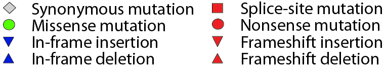
Figure notes
• "Mouse over" a mutation to see details. |
|
Click on a tumor type to see its full list of significant genes.
Data details
 TumorPortal
 Sign in